
Three standard ways of measuring a person’s capacity to talk themselves out of a bad feeling agree with each other almost not at all, according to a consortium analysis of 2,175 people across 40 brain-imaging studies, published in Nature on October 28. The strongest of the three pairwise relationships, between how much relief people reported during a lab task and how much their amygdala activity dropped on a brain scan, came out small enough that a single study would need 1,105 participants to detect it reliably at 80% statistical power — a sample size almost no fMRI study has ever approached, as Ars Technica’s John Timmer wrote of the paper. The questionnaire that asks people how often they reframe their problems in daily life didn’t correlate with either of the other two measures at all.
The skill in question is cognitive reappraisal: taking a situation that triggered a negative emotion and finding a different way to interpret it so it stings less. It’s the mechanism cognitive behavioral therapy is built around, and it’s been linked in decades of smaller studies to emotional resilience and lower symptom burden. The problem the new paper raises is that researchers have been using three different instruments to measure this one skill, on the assumption that they’re all tapping the same underlying thing. They aren’t.
Three instruments, one assumed target
The first instrument is a trait questionnaire: a self-report asking how often, in general, someone reframes stressful situations. It covers a long time horizon and inevitably picks up personality traits well beyond emotion control. The second is a task-based rating: inside an MRI scanner, a participant views a negative image, is told to either just look at it or to reappraise it, and then rates in the moment how they feel. The third is the amygdala itself — a brain structure that activates during emotional responses — measured by how much its blood-oxygen signal drops when someone reappraises versus when they just look. The assumption behind thirty years of affective-neuroscience papers is that someone who scores high on the questionnaire should also show bigger in-scanner mood shifts and bigger amygdala drops. The consortium’s data say no.
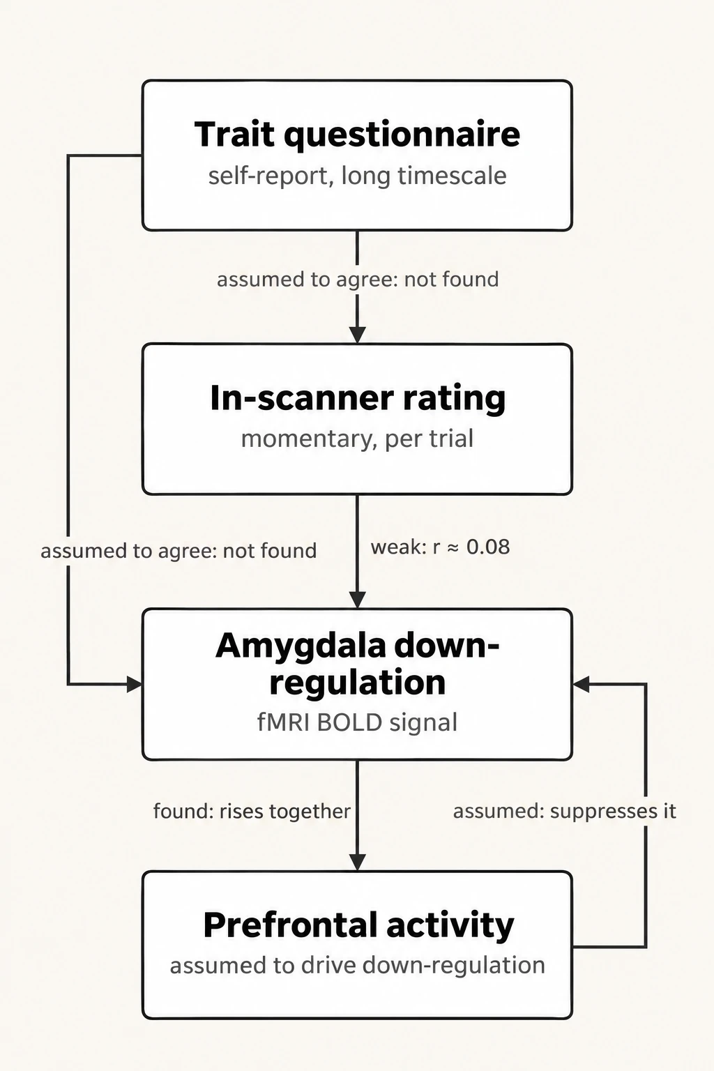
| From | To | How |
|---|---|---|
| Trait questionnaire (self-report, long timescale) | In-scanner rating (momentary, per trial) | assumed to agree: not found |
| Trait questionnaire (self-report, long timescale) | Amygdala down-regulation (fMRI BOLD signal) | assumed to agree: not found |
| In-scanner rating (momentary, per trial) | Amygdala down-regulation (fMRI BOLD signal) | weak: r ≈ 0.08 |
| Prefrontal activity (assumed to drive down-regulation) | Amygdala down-regulation (fMRI BOLD signal) | assumed: suppresses it |
| Amygdala down-regulation (fMRI BOLD signal) | Prefrontal activity (assumed to drive down-regulation) | found: rises together |
Based on NIDER consortium / Nature
That r ≈ 0.08 figure is the paper’s single strongest cross-measure correlation, and it means the two measures share under 1% of their variance. Put differently: knowing how much someone’s mood improved after reappraising inside a scanner tells you almost nothing about how much their amygdala activity dropped during the same trial, even though both are supposed to be reading out the same underlying capacity.
The scale of the check
The analysis behind this came from NIDER — the Neurobiology of Individual Differences in Emotion Regulation consortium — in which 32 research teams pooled 40 fMRI datasets, nine of them previously unpublished, for a combined sample of 2,175 participants, according to a press release from Germany’s Central Institute for Mental Health, whose researcher Maurizio Sicorello led the project with Carmen Morawetz, professor of affective neuroscience at the University of Innsbruck. That sample is roughly 20 times larger than the biggest single cohort in a prior review of this literature, which is the scale needed to detect small effects reliably rather than mistake noise in a 20- or 30-person study for a real signal — the typical size of past reappraisal fMRI work.
A second, separate finding complicates the field’s usual story about mechanism. The standard model holds that prefrontal cortex regions, responsible for higher-level reasoning, activate during reappraisal and suppress the amygdala — more prefrontal activity, more amygdala down-regulation, in opposite directions. The consortium’s data show the two regions moving together instead: participants with stronger amygdala responses during reappraisal also tended to show stronger activity elsewhere in reasoning-related cortex, and weaker amygdala responses went with weaker activity there too. That’s the correlation running the same direction, not the inverse relationship the suppression model predicts. The paper also found that people’s own belief in how well they reappraise correlated with amygdala down-regulation in the wrong direction — those who thought they reframed well showed less amygdala change, not more.
This doesn’t mean the prefrontal cortex is unimportant for emotion regulation. For our field, this is an uncomfortable but important insight.
That’s Morawetz, quoted in the Central Institute’s release. Her point is narrower than it might sound: brain activity, she said, can still reveal how an individual’s own emotions change over time within that person. What it can’t do, on this evidence, is stand in as a cross-sectional biomarker that ranks people against each other — the use case most clinical neuroscience papers actually rely on, including studies that treat lower prefrontal activity in patients as evidence of impaired emotion regulation. Sicorello, in the same release, framed the underlying skill as less like a trainable muscle and more a product of habitual thinking patterns: whether someone looks for alternative interpretations of a bad situation in the first place, and whether an alternative interpretation feels credible to them once they find one.

The consortium’s alternative candidate regions — slightly reduced activity in sensorimotor and attention networks among people who regulated more effectively, plus some signal in the amygdala and brainstem — are regions more associated with generating emotional responses than regulating them, and the paper itself describes these effects as small and sensitive to which datasets are included, not a replacement biomarker ready to use.
What this leaves unmeasured
None of this proves reappraisal capacity isn’t real; CBT’s clinical track record doesn’t rest on amygdala scans. It does mean that three decades of papers linking prefrontal-amygdala coupling to individual differences in emotion regulation were built on an assumption — that self-report, in-scanner behavior and neural signal converge on one construct — that a sample 20 times larger than anything before it failed to confirm. The paper’s own caution, that strong claims about a person’s emotion-regulation capacity based on any single brain region should be made with considerable caution, is aimed at a decade of smaller studies that made exactly that claim. Morawetz’s stated fix is to build more realistic, personally relevant tasks rather than the simplified view-versus-reappraise paradigm used across most of these 40 datasets. Whether a redesigned task, run at anything like NIDER’s scale, would finally produce measures that agree with each other is the next number this field owes someone 1,105 participants’ worth of data to answer.