
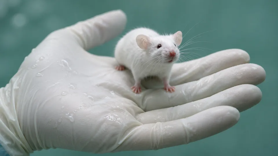
The cortex on the cover of Nature on Oct. 8 belonged to a mouse, and 91.9 percent of it, by volume, was human.
That figure comes from a Stanford paper published online Sept. 16, which describes 25 animals whose own neocortex was engineered never to form and whose empty forebrain was then filled with lab-grown human cortical tissue. The human cells grew on a human clock, sent axons into the host’s brain stem and cervical spinal cord, and produced a neuron type that no culture dish has ever yielded.
The scientific claim here is narrower than the headlines and the regulatory claim is wider. The model’s two advertised uses, frontotemporal dementia and psychiatric illness, rest on very different amounts of evidence, and only one of them is close. Meanwhile the line that stops this work short of monkeys is drawn by the people doing it. Under the main international guidelines, putting human neural cells into the brain of an animal that has already been born falls in the lowest tier of oversight, exempt from any specialized ethics review. The federal funding moratorium usually invoked in these arguments applies only to embryos. Two state laws that criminalize human-animal hybrids with mostly human brains both carve out the exact procedure Stanford used.
What the graft did
The mice start out missing something. By deleting Esco2 in the pallial cells that would become neocortex and hippocampus, the lab led by Sergiu Pașca, a professor of psychiatry and behavioral sciences at Stanford, produced what it calls apallial mice: animals with about 2 percent of the normal cortical content and roughly half the usual brain volume on M.R.I. Dr. Pașca is insistent that nothing was taken out. “This is not a depletion. It’s not an ablation,” he told Inside Precision Medicine. “This is sort of like a prevention of the formation. It’s a blockade.”
Into that cavity went up to four human cortical organoids per animal, each about 100,000 cells and 30 to 60 days old, grown from induced pluripotent stem cells donated by healthy people who had consented to their cells being put into animals. Accounts of the recipients’ age differ: Stanford’s own announcement and Al Jazeera describe 2-day-old pups, while a summary of the paper’s methods by News-Medical gives hosts of 5 to 17 days.
Twenty-nine mice got the surgery, with organoids from three stem-cell lines. Grafts survived in 86.2 percent of them. Between the second and third month the human tissue grew 4.7-fold, tapping the mouse circulation, and at three months it accounted for 91.9 percent of the combined cortical volume. Some coverage renders this as 92 percent of the cells in the rebuilt cortex, which is a different measurement; and across the whole brain, human tissue came to roughly half the volume, not nine-tenths.
The authors reported 880,149 single-nucleus profiles from the grafts. They found more than three times the proportion of layer 5 extratelencephalic projection neurons seen in earlier transplant work, calcium bursts lasting tens of seconds that recurred every few minutes and spread locally across the graft in about 100 milliseconds, and, in all three animals sampled for it, cells with the molecular signature and long spindle shape of von Economo neurons.
It is not a cortex. There are no layers. “There’s no lamination in the human graft in the xenocortical animals,” Dr. Pașca said, adding that the cells “don’t know where up and down really is.” Inhibitory interneurons are scarce. At 24 weeks after differentiation the grafted glutamatergic neurons matched mid-gestation human cortex; NPR reported that they resemble a third-trimester fetus. Either way, this is fetal tissue in an adult animal, and Dr. Pașca told NPR that human neurons develop at least 20 times more slowly than mouse ones.
The behavior was mixed, and the paper says so. On a Y-maze working-memory task, normal mice and grafted mice scored above chance and apallial mice did not; the authors note the Y-maze result does not establish that the human graft restored working memory, that anatomical connectivity “does not demonstrate pathway-specific functional integration,” and that it is unclear whether graft neurons are “necessary or sufficient for particular behaviors.” Five hours at 5 percent oxygen damaged the human tissue and left the animals unsteady, which normal and apallial mice were not.
Today in @Nature, we report a new approach to studying human brain development and modeling disease in vivo.
Human neural #assembloids and #organoids have opened new ways to study development in vitro, but they also have important limitations. Transplantation can provide a more…
The dementia case
Von Economo neurons are the strongest card in the deck, and they are strong for a specific reason. Described by Constantin von Economo and Georg Koskinas in 1925, they sit in layer 5 of the anterior cingulate and frontoinsular cortex, appear only in humans, apes, elephants and cetaceans, and amount to roughly one in 90,000 cortical neurons. In 2006 William Seeley and colleagues at the University of California, San Francisco, found selective loss of these cells in mild to moderate behavioral variant frontotemporal dementia but not in Alzheimer’s disease. A 2012 study in Cerebral Cortex by Eun-Joo Kim and the same group quantified the loss in the frontoinsula and found the cells dying off while neighboring layer 5 neurons survived.
Mice have no von Economo neurons to lose. Organoids grown in a dish do not make them. The xenocortical grafts did, which is the single result here that neither of the two existing model systems can produce at all.
Dr. Pașca has named the experiment he wants to run: derive cortical tissue from people with frontotemporal dementia, including carriers of defined causal mutations, put it in these mice, and watch which cells go first.
One unique application of this is the study of von Economo neurons in the context of frontotemporal dementia.
There is a catch, and it is the same catch that has dogged organoids. Reprogramming a skin cell to a stem cell wipes out epigenetic age, telomere shortening and accumulated DNA damage. A 2024 review in Frontiers in Cellular Neuroscience argues that organoids cannot activate an aging program at all and are therefore unreliable models of the aging human brain. Nothing in the xenocortication method fixes that. The graft is on a human developmental clock, which is the point, but a human developmental clock run for six months reaches mid-gestation, not 60. What the model can plausibly show is cell-intrinsic vulnerability in a rare neuron type, inside a living circulatory system, with a behaving animal attached. It cannot show late-onset degeneration.
The schizophrenia case
Here the argument is weaker, and the history explains why. The pitch, as Stanford put it, is that the method should speed work on schizophrenia, epilepsy, profound autism and cerebral palsy, because the implanted cells carry the donor’s genome and because, in Dr. Pașca’s words, circuit and behavioral readouts matter for neuropsychiatric disorders in a way they do not for a dish. He has made the sharper version of the point too: a neuron that is hyperexcitable in culture has not thereby caused a seizure.
Chimeric mice have already been pushed at schizophrenia once. In 2017 Martha Windrem, Steven Goldman and colleagues at the University of Rochester transplanted glial progenitors derived from the iPS cells of patients with childhood-onset schizophrenia into mice, and reported in Cell Stem Cell delayed astrocytic differentiation, hypomyelination, reduced prepulse inhibition, excessive anxiety, antisocial behavior and disturbed sleep. That was nine years ago, with glia rather than neurons, and it has not produced a treatment.
The deeper objection is genetic. Writing in Philosophical Transactions of the Royal Society B in 2018, Steven Hyman of the Broad Institute concluded that because polygenic combinations of alleles underlie psychiatric risk, and because poorly conserved noncoding regions hold most of the common disease-associated loci, “the already distant possibility of a genetic mouse model of schizophrenia or a mood disorder fades to impossibility.” Xenocortication answers part of that, since a patient-derived graft carries the whole polygenic background rather than one edited gene. It does not answer the timing problem: schizophrenia declares itself in late adolescence, and the graft never gets there.
Joao L. de Quevedo, who directs the Center for Interventional Psychiatry at UTHealth Houston, wrote in October that patient-derived variants in living circuits are a possibility for future experiments “not diseases shown to be reproduced or treated in this study,” and that the work tested no psychiatric intervention of any kind. On the record, the dementia application has a result behind it. The schizophrenia application has a rationale.
Earlier chimeras
This is the fourth generation of the same idea. In March 2013, Xiaoning Han, Dr. Goldman and Maiken Nedergaard [reported in Cell Stem Cell](https://www.cell.com/cell-stem-cell/fulltext/S1934-5909(13)00007-6) that human glial progenitors engrafted into newborn mice outcompeted the host’s own glia, sharply enhanced long-term potentiation, and improved learning across four tasks, including the Barnes maze and fear conditioning. Mice given mouse glia showed no such gain. Human astrocytes, in other words, made mice measurably better learners 13 years ago, and the result drew a fraction of this year’s attention.
In 2019, Daniele Linaro and Pierre Vanderhaeghen’s group transplanted individual human cortical neurons into neonatal mouse visual cortex and found that they kept their own slow developmental schedule while eventually responding to visual stimuli like their mouse neighbors. Mouse cortical neurons mature in three to four weeks, macaque neurons in three to four months, human neurons over years. That mismatch is what Dr. Pașca’s 2022 rat experiment ran into, and what clearing the cortex was designed to defeat. “While we create more space, we still don’t solve the problem of time,” he said.

Where the rules stop
The oversight question has a clean answer, and it is not the one most readers would guess. The 2021 chimera guidelines of the International Society for Stem Cell Research, written by a subcommittee chaired by the bioethicist Insoo Hyun and published in Stem Cell Reports, split the field by timing. Human cells introduced into an animal embryo or fetus get Category 2: permissible only after specialized scientific and ethics review. Human cells introduced after birth get Category 1A.
Falling under category 1A is the transfer of human stem cells, their derivatives, or other human cells into postnatal animal hosts.
Category 1A means exempt from specialized review, handled by the ordinary institutional animal-care committee, supplemented by stem-cell expertise for work in the central nervous system. The same subcommittee held that scenarios such as laboratory animals with human-like cognitive traits are “currently scientifically out of reach,” and warned against confusing structural humanization with moral humanization, noting that such traits “are not biologically assured even in infant brains that are 100% human.” The 2025 revision left those provisions in place.
The federal picture is thinner still. The N.I.H. moratorium of Sept. 23, 2015 covers human pluripotent cells put into pre-gastrulation animal embryos, which is not what happened here. The agency’s August 2016 proposal to lift it and route sensitive cases to an internal steering committee drew more than 22,000 public comments, according to the Animal Legal Defense Fund, and was never finalized; Undark reported in September that the moratorium remains in force. The National Academies said the rest of it plainly in April 2021.
there are no provisions in U.S. federal law specific to the generation of human-animal chimeras or to ensuring their welfare and well-being
Louisiana and Arizona do criminalize creating a life form whose brain is derived wholly or predominantly from human neural tissue. Nita Farahany of Duke and Anil Seth of the University of Sussex point out in the Journal of Law and the Biosciences that both statutes exempt postnatal xenotransplantation, the category Stanford’s work falls into. Britain, by contrast, requires national review for substantial modification of an animal’s brain that may make its function potentially more human-like.
So the binding constraint on this experiment was Stanford’s own: prospective consultation with university bioethicists, review by the Stanford Neuroscience Institute executive committee, the stem-cell research oversight committee, the animal-care committee, and an independent ad hoc ethics board that included outside neuroscientists, legal scholars and patient advocates. Dr. Farahany sat on that external board, unpaid. One of the limits it helped set was stopping at six months.
The objections weighed
Asked by MIT Technology Review where he would not go, Dr. Pașca named one species.
One of the things that I see as a very clear red line is doing this experiment in a primate.
The paper itself is less categorical. Its discussion names cortically depleted embryonic rodent hosts, or non-human primates, as routes to reducing the developmental mismatch between graft and host. A red line in an interview and a listed future direction in the same document are not the same position, and the record does not say which one governs the lab’s next grant.
John Evans, a sociology professor at the University of California, San Diego, who was not involved, told Al Jazeera that the mouse work raises no major ethical problem on its own terms but demonstrates the path to animals that would. His objection is structural: between a mouse and a macaque there is no defined stopping point other than a researcher’s judgment. The documents bear him out. Category 1A does not scale with graft size, host species, or survival time.
Julian Koplin of the University of Melbourne made the moral-status version of the argument in Bioethics in 2022, writing that the ISSCR recommendations “fail to address the core ethical concerns raised by neurological chimeras” and rest on philosophical claims “too tenuous and controversial to underpin global guidelines.” He also allows the other side of it: given the potential benefits, he writes, there is a moral imperative not to restrict the work without good reason.

The consciousness worry is the one the evidence currently handles worst for the people raising it. Dr. Farahany and Dr. Seth, asking what it is like to be a xenocortical mouse, answer that as things stand it may well be nothing at all, and compare the graft’s slow cortex-wide waves to an orchestra tuning up rather than performing. The trace-conditioning deficit that has been linked to consciousness in animals is ambiguous here, they note, because apallial mice showed the same deficit, so the assay may be tracking the missing mouse cortex rather than the human graft. Sarah Chan, a reader in bioethics at the University of Edinburgh, told BBC News there was no indication the work produces mice that think like humans.
The objection that the record does support is smaller and more awkward. Mackenzie Graham, a senior research fellow at the University of Oxford, argued in The Conversation on Oct. 19 that welfare assessment in laboratory animals is inference from behavior, and that rebuilding the organ that generates both the experience and the behavior breaks the inference. Reading reduced grooming as distress assumes grooming still means what it meant. Nothing in the Stanford paper validates that assumption, and nothing in Category 1A requires anyone to.
Long before we create an animal that thinks like us, we may create one whose behaviour we no longer understand.
James Ainge, a neuroscientist at the University of St Andrews, told BBC News the work was technically impressive but that the mice could be of limited use, partly because of the ethical constraints on housing living human brain tissue in animals. His colleague Ilary Allodi described the scans as a bit messy, and the graft as the human program running inside a mouse incubator.
What comes next
Dr. Seth announced the commentary on X the day the paper appeared, calling the result a milestone in chimera research and promising a roadmap with Dr. Farahany.
An extraordinary @Nature paper has just landed from the lab of @Sergiu_P_Pasca at @Stanford - led by Konstantin Kaganovsky. They have found a way to create “xenocortical mice” which have ~92% of their cortex occupied by a cortical organoid derived from human stem cells. This…
That roadmap, published in the Journal of Law and the Biosciences, proposes no bans. It asks for staged escalation with review checkpoints fixed in advance; mandatory independent review before crossing a threshold such as longer maturation, assembloid grafts, embryonic hosts or a new species; a pre-transplantation protocol record and interim welfare reports to committees empowered to pause or terminate a study; donor consent specific to in vivo use rather than blanket consent; and a reversal of the burden of proof on host species.
Nature ran an editorial on Sept. 29 calling for international oversight of lab-grown brain organoids, having argued in an earlier editorial that the concerns do not warrant a moratorium. China’s National Science and Technology Ethics Committee issued binding human organoid research guidelines on April 29, 2025, the first national framework to cover brain organoids, organoid chimeras and embryo models together; the ISSCR has a revision of its own guidelines in progress.
Dr. Pașca’s lab has trained close to 500 laboratories in organoid generation, and he says the Emx1-Cre mice the method depends on already exist. “We built the system so others can also use it,” he said. “We’re not going to be able to do everything.” Stanford’s Office of Technology Licensing holds a provisional patent application on organoid transplantation, with Dr. Pașca and four co-authors named as inventors.
The six-month ceiling was a choice, not a finding. Whoever runs the experiment at 12 months will not need a specialized ethics review to do it.